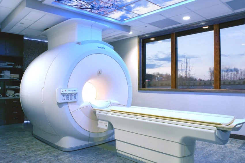
3 TESLA MRI MACHINE
MRI, or magnetic resonance imaging, uses magnets and radio waves to provide images of the internal structures of the body. Magnetic resonance imaging allows medical professionals to examine the inside of a person’s body without the need for an invasive procedure. Unlike x-rays, MRI uses non-ionizing radiation. A 3-tesla magnetic field is twice as powerful as the fields used in conventional high-field MRI scanners, and as much as 15 times stronger than low-field or open MRI scanners. This results in a clearer and more complete image. Stronger magnetic fields also produce better images of soft tissues and organs than standard MRI scanners. 3T MRI is the fastest diagnostic magnetic resonance imaging technology available. The computer generated images can be sent off-site for immediate analysis. These exams can be up to 100 times faster than standard MRI exams. Another advantage of 3 Tesla MRI is that the exams offer more comfort than typical MRI exams. Some patients may feel anxious or claustrophobic in traditional MRI scanners. 3 Tesla MRI offers a more spacious bore, or tube, and the patient’s head can remain outside of the bore for scans that don’t involve the spinal cord, neck, or head.
THE ADDITIONAL ADVANCED PROTOCOLS WE PROVIDE
FUNCTIONAL MRI
MRI, or magnetic resonance imaging, uses magnets and radio waves to provide images of the internal structures of the body. Magnetic resonance imaging allows medical professionals to examine the inside of a person’s body without the need for an invasive procedure. Unlike x-rays, MRI uses non-ionizing radiation. A 3-tesla magnetic field is twice as powerful as the fields used in conventional high-field MRI scanners, and as much as 15 times stronger than low-field or open MRI scanners. This results in a clearer and more complete image. Stronger magnetic fields also produce better images of soft tissues and organs than standard MRI scanners. 3T MRI is the fastest diagnostic magnetic resonance imaging technology available. The computer generated images can be sent off-site for immediate analysis. These exams can be up to 100 times faster than standard MRI exams. Another advantage of 3 Tesla MRI is that the exams offer more comfort than typical MRI exams. Some patients may feel anxious or claustrophobic in traditional MRI scanners. 3 Tesla MRI offers a more spacious bore, or tube, and the patient’s head can remain outside of the bore for scans that don’t involve the spinal cord, neck, or head.

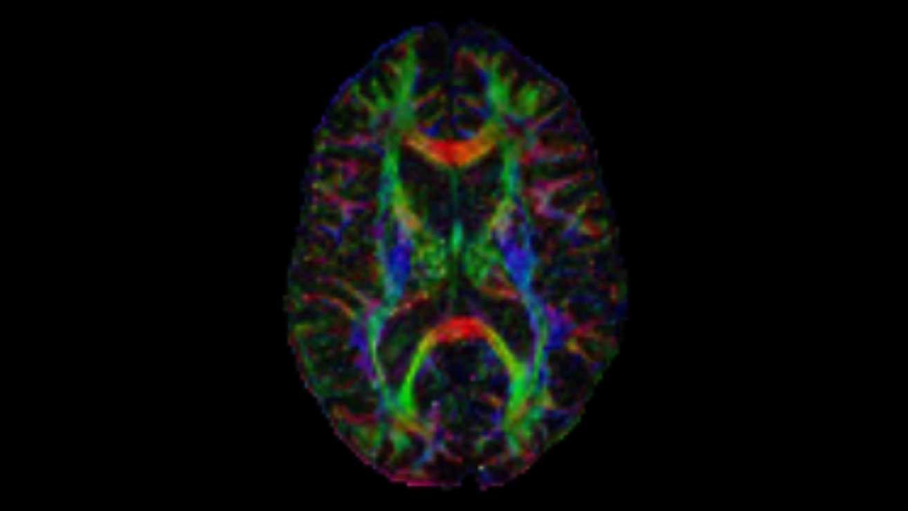
DIFFUSION TENSOR IMAGING
The architecture of the axons in parallel bundles and their myelin shield facilitate the diffusion of the water molecules along their main direction. If we apply diffusion gradients in at least 6 non-collinear directions, it is possible to calculate, for each pixel, a diffusion tensor (i.e. a 3*3 matrix) that describes this diffusion anisotropy.
The fiber’s direction is indicated by the tensor’s main eigenvector. This vector can be color-coded, yielding a cartography of the tracts’ position, direction (red for right-left, blue for foot-head, green for anterior-posterior), and anisotropy (as indicated by the tract’s brightness). The brain’s main white matter tracts can be recognized (White Matter Atlas).
In addition, the apparent diffusion coefficient (ADC) and fractional anisotropy (FA) can be quantified.
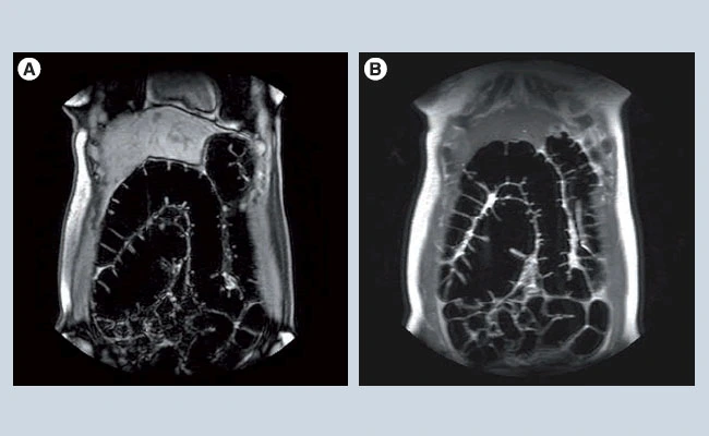
MR COLONGRAPHY
Magnetic resonance imaging of the colon is widely used for the diagnosis and treatment of anorectal disorders, but other applications can be considered as well. In this article we discuss the application of MRI in colorectal cancer, inflammatory bowel disease (Crohn’s disease and ulcerative colitis) and acute abdominal pain. The latter concerns MRI without a specific preparation while the other two applications concern the use of bowel preparation and colon distension (magnetic resonance colonography). CT is now the principal technique for these applications. The major advantage of MRI of the colon over CT is the lack of ionizing radiation exposure. The high soft-tissue contrast might be another advantage. Research over the last decade has demonstrated that MRI is a valuable alternative to CT, with accuracy values equal or superior to CT. Colorectal diffusion-weighted imaging has been increasingly studied with promising results.
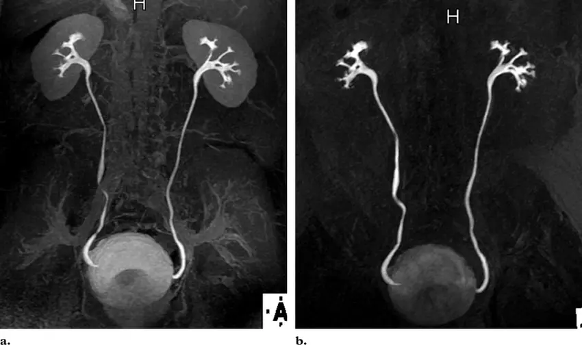
MR UROGRAPHY
Magnetic resonance urography (MRU) is a radiation-free exam that uses magnetic waves to create detailed pictures of the kidneys, ureters and bladder. It is a radiation-free way to look at the structure and function of the urinary tract, which is the part of the body that produces and transports urine.
The MRU study is done in a magnetic resonance imaging (MRI) machine. A urinary catheter (tube inserted through the urethra and into the bladder) and an intravenous (IV) catheter (tube inserted through the skin into the vein) are required. Together, these catheters help us measure drainage of the urine to assess the function of your child’s kidneys. The MRU study takes about one hour.
OTHER SERVICES
* Management and Quality Improvement systems for Hospital and Private Clinical Laboratories
* Developing Quality Systems according to the guidance of US Accreditation agencies such as CAP (College of American Pathologists), CLSI (Clinical Laboratory Standards Institute)

* Developing Standard Operating Procedures for Clinical and Histopathology Laboratories
* Laboratory Guidelines and Operating Procedures by using US Standards
* Information Technology in health and other related industries
* Training Laboratory Scientists/Technicians
* Training mid- level managers for laboratory, nurses and IT administration
* Quality Assurance and Improvement and maintaining proficiency testing under the guidelines of CAP
* Providing education and training for Physicians conducting complicated cases and implantation procedures
* Clinical Pathology consultation, diagnosis, interpretation and reporting systems
* Equipment and Software validation for Hospital, private clinical laboratory, and reference laboratory
* Our team of experts can provide guidance for developing and operating any clinical laboratory, Stem cell clinics, and doctor’s offices
SERVICES ACCREDITED BY JCAHO
Ensuring Joint Commission regulations
Ensuring to follow Env of care standards-
EC.02.01.xx- Safety & security
EC.02.02.xx- Hazmat & waste
EC.02.03.xx- Fire Safety
EC.02.04.xx- Medical Equipment
EC.02.05.xx- Utilities
EC.02.06.xx- Other Physical Environments Requirements

Taking care of Sentinel Event Alerts- Equipment related alerts
1. SEA 38: MRI safety
2. SEA 36: Tubing misconnections
3. SEA 27: Bed rail entrapment
4. SEA 25: Ventilator alarms
5. SEA 21: Medical gas mix-ups
6. SEA 15: Infusion pump free-flow
Ensuring NFPA 99 standards are followed for Health care Facilities
10.5.2.1.2
8.4.1.3.5 Chassis Leakage current
8.7.3 Touch Current
8.4.1.3.2 Ground conductor resistance
8.6.4 Protective Earth Impedance
* Ensuring to follow FDA regulations for Medical Equipment
* Ensuring FCC (Federal Communications Commission) guidelines
* Ensuring National Patient Safety Goals (NPSG)